Stories and Style in Early Modern Anatomical Illustration
By Tracy Molis•August 2023•11 Minute Read
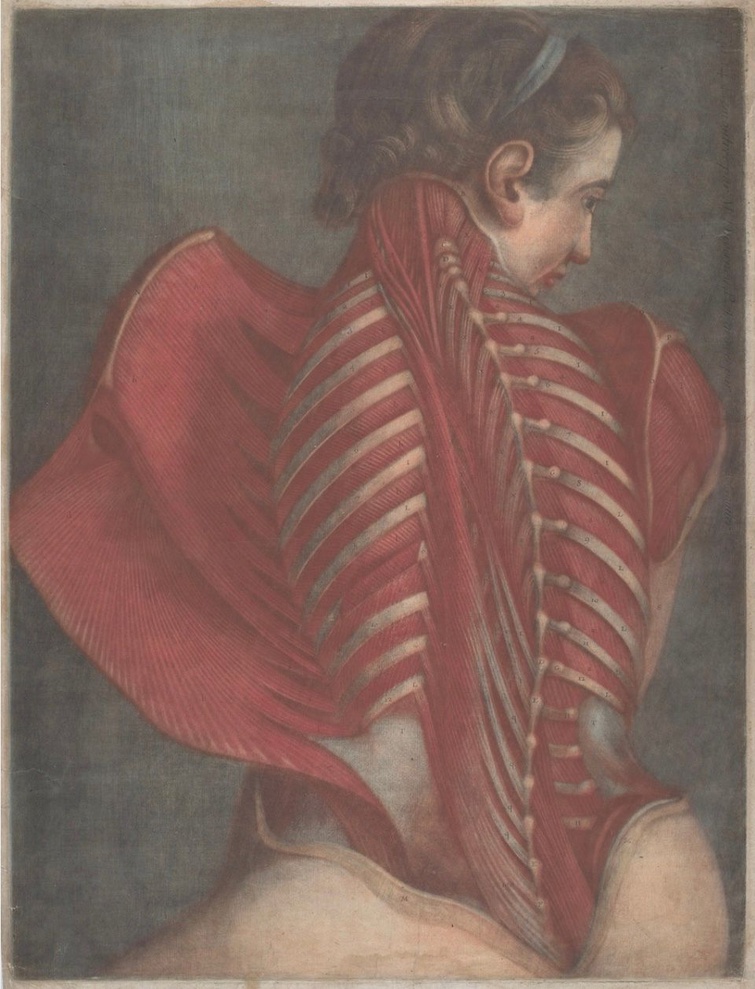
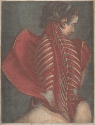
Jacques-Fabien Gautier Dagoty, L'Ange anatomique (The Anatomical Angel), or Dissection of a Woman’s Back, 1746. National Gallery of Art, public domain.
In the early modern period, anatomists and illustrators teamed up to create the most comprehensive guides on human anatomy then available. Examining the visual styles and symbolism of these anatomical illustrations can reveal a glimpse into their viewpoints around the body that still reverberate today.
In Renaissance and Enlightenment Europe, anatomists who explored the human body in depth faced complex stylistic choices when deciding how to portray their observations visually. Well before the standardization of medical textbooks, anatomist-illustrators looked to tropes from antiquity as models for developing these images. They often addressed moral questions around death, birth, and humanity's place under Christianity. Dissecting the body was not solely the domain of medicine, but also touched upon the realms of philosophy and theology. Some doctors of the era dramatized their pursuits through the iconic image of the operating theater.
Much like maps, anatomical atlases take on an imperialist or "God's eye" view in order to simplify complex and confounding natural phenomena into diagrams legible to their intended audience. Whether we find them beautiful or disturbing, anatomical images and the rhetoric they encode in their aesthetic styles reveal more about the values of the time period than the objective reality of, as it were, the terrain.
The Heroic Style
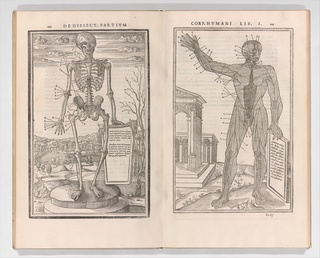
A series of flayed men flex their exposed muscles in picturesque Hellenic landscapes. Marble-like torsos display open abdomens, revealing intricately detailed viscera. One with breasts sports a flowing lock of hair. When Andreas Vesalius first published De Humani Corporis Fabrica Libri Septem in 1543, he employed acolytes of the painter Titian to illustrate Europe's first compendium of human anatomy. This is one reason why these woodcuts may appear more like melodramatic Renaissance artworks than the rigorous science guides they were intended to be.
Andreas Vesalius & Jan Steven van Calcar, De humani corporis fabrica (Of the Structure of the Human Body), 1555. Woodcut. The Metropolitan Museum of Art, public domain.
However, Vesalius's Fabrica was the leading edge of empiricism at the time. He revolutionized medical instruction through cadaver dissections in European medicine. Previously, an instructor would lecture from a classical volume by the ancient Greek anatomist Galen, then have a barber-surgeon cut apart an animal for the class to view, believing they were similar enough in structure to demonstrate the human form. This was similar to Galen’s research, as he wrote his treatise by examining the anatomy of pigs, oxen, or Barbary macaques. Vesalius turned that on its head by advocating for direct knowledge and observation of human corpses, refuting many of Galen's earlier claims in the process. He created the Fabrica to serve as a more objective guide for Renaissance physicians than what was previously available.
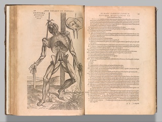
The notion of an "objective" style in Western scientific imagery changed throughout the centuries until the 1850's, when medical illustration moved toward the so-called "style-less" Gray's Anatomy mode of diagrams we are most familiar with today. In the 16th century, however, anatomists like Vesalius framed their practice in light of the dictum nosce te ipsum—know thyself. For them, uncovering the body's structure and mechanisms was inseparable from discovering humanity's essence. It was a heroic philosophical pursuit deserving of a heroic aesthetic. Charles Estienne, who published his De dissectione partium corporis humani libri tres in 1545, set out to create a grand story of the body going back to Galen. Although these early medical books referenced pagan antiquity, they stood firmly within a monotheist Christian tradition. The composition of Estienne's picture demonstrating the nervous system referenced creationism. We see an electric-looking figure, nerves twitching, suspended like hairs on end. He writes that the human nerves resonate with the “concordance and harmony” of universal design.1
Charles Estienne, De dissectione partium corporis humani libri tres, 1545. The Metropolitan Museum of Art, public domain.
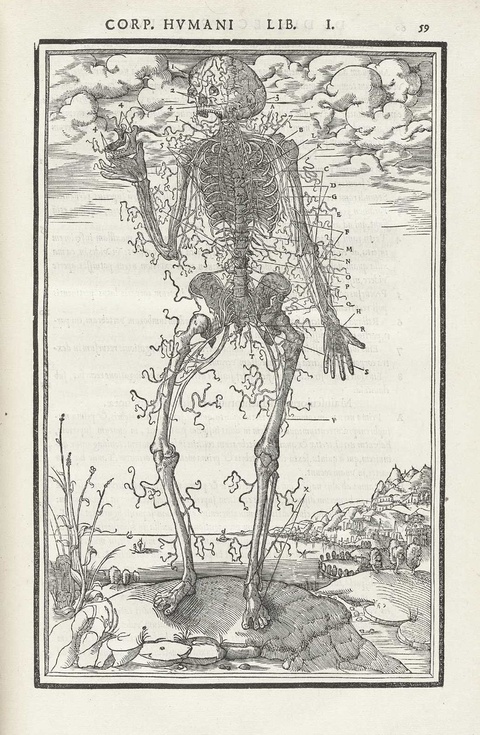
Mortality was a common theme in these compendiums. Vesalius had his artists inscribe a quotation from Virgil on an illustration of a classical plinth, upon which a skeleton solemnly contemplates a skull: "Genius lives on, all the rest will perish." The lofty visual rhetoric of these compositions addressed an uncomfortable fact. To Renaissance Christians, dissecting cadavers was carving up God's highest creation. It was appropriate that their illustrations reflected the handiwork of the divine. Although they mostly sourced bodies from executed criminals, for whom the use of their bodies was part of the punishment, the authors of these compilations were well aware of the fraught moral line they trod.2
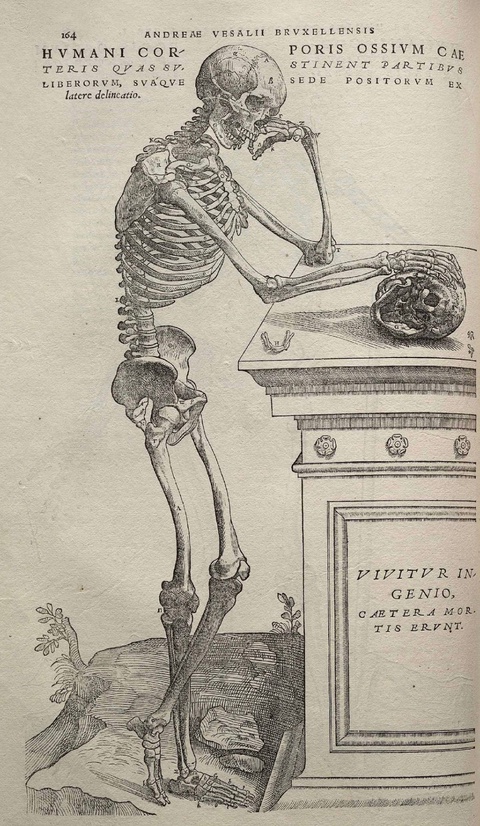
Andreas Vesalius & Jan Steven van Calcar, “Skeleton,” from De humani corporis fabrica (Of the Structure of the Human Body), 1543, pp. 164. Woodcut. Wikimedia commons, public domain.
Eighteenth century advancements in printing technologies allowed more refined, larger format books than the woodcut types of the early Renaissance. Illustrators like Jacques-Fabien Gautier d'Agoty skillfully produced editions in full color using new methods with mezzotint plates, as we can see in his Dissection of a Woman's Back, from 1746.3 The work's alternate title, The Anatomical Angel, alludes to the Christian afterlife while giving it the connotations of a striking piece of art.
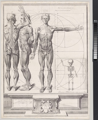
Idealized illustrations representing the beauty of "God's handiwork" culminated in the 18th century.4 Another example is Musculorum Tabula IV illustrated by Jan Wandelaar from Tabulae Sceleti et Musculorum Corporis Humani in 1747. The author of this volume, Bernard Siegfried Albinus, touted the extreme effort they devoted to these precise renderings in order to synthesize—from many sources—their notion of the most "perfect" and "just" specimen of a skeleton.5
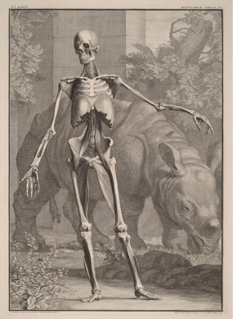
Bernard Siegfried Albinus & Jan Wandelaar, Musculorum Tabula IV, 1742, from the series Tabulae Sceleti et Musculorum Corporis Humani. Engraving on laid paper. National Gallery of Art, public domain.
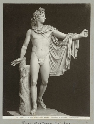
In this view, one could only obtain scientific objectivity by creating ideals, rather than representing the imperfect variations of a type within individuals. The result is a completely smooth anatomical model that resembles polished ivory. He stands in the pose of the Apollo Belvedere with his hand lording over a rhinoceros in the background. Why a rhinoceros? Albinus states it was an "ornament" they chose for "the rarity of the beast." The pose also symbolizes the hierarchy of "man" over "nature" in both the Bible and the Humanist tradition.
Trompe l'oeil and Realism
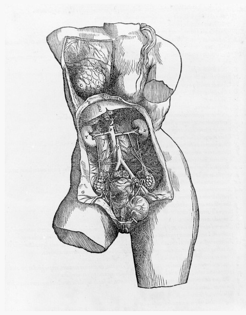
A separate strain of thought developed out of Vesalius's commitment to close observation, represented by the picture in the Fabrica of all his tools laid out starkly on his vivisection board. Some anatomists set out to remedy stylistic excesses by portraying individual specimens with hyper-realistic accuracy. These illustrations worked hard to convince the viewer they were seeing the unvarnished, most typical example, in what we can call "the rhetoric of the real."6 They hearkened back to the ancient tradition of trompe l'oeil by depicting a window's reflection on a membrane or a fly landing on the fabric above the cadaver.
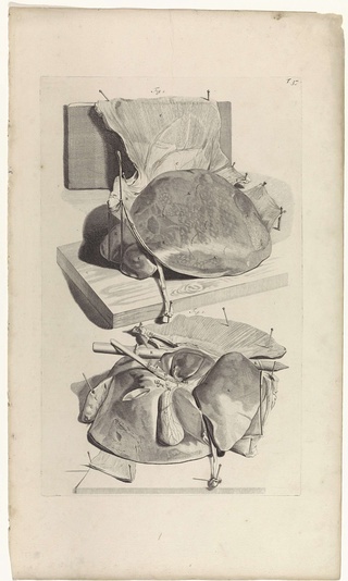
An early book in this style was Govard Bidloo's Anatomia humani corporis with renderings by Gérard de Lairesse, from 1685. The images included the accessories of dissection, as if to prove the truth of the scene. However, the graphic nature of this style risked presenting the viewer with mess and confusion. It did little to clarify the structures one was looking at, or how they fit in the context of the body. Lairesse's depiction of the liver, stuck with pins on the cutting board, makes it apparent why some degree of stylistic simplification became necessary for anatomical illustrations to function as practical guides.7
Anatomy Theater
Many of the aforementioned physicians didn't limit rhetorical flair to the instructional parts of their books. Vesalius started performing public human dissections in 1543 in Basel, Switzerland, dramatizing the practice in the frontispiece of his Fabrica.
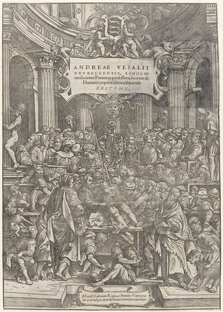
In this depiction, spectators clamor in a grandiose temple to watch him cut into a body’s uterus. This self-mythologizing image relied on the Renaissance cult of the male genius, interpreting the female reproductive organs to reveal the origins of life itself.8 The trope of the dissection theater proliferated in the popular consciousness as the practice evolved into official surgeon’s guild events open to the general public.
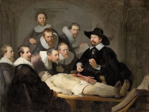
Rembrandt van Rijn, The Anatomy Lesson of Dr. Nicolaes Tulp, 1632. Oil on canvas. The Mauritshuis Museum, public domain.
Rembrandt's painting The Anatomy Lesson of Dr. Nicolaes Tulp of 1632 is a well known example. Some features of this scene clue us in to Rembrandt's iconographic intentions. According to protocols of the time, the doctor would start by dissecting the soft organs of the chest and thorax. By leaving this part of the male figure intact, Rembrandt gives the corpse a Christlike appearance, and the composition's focus goes to the forearm. Dr. Tulp holds up the muscles responsible for the hand's fine motor control in his forceps. As one of the foremost scholars on primates in his day, Tulp would likely have drawn attention to how the opposable thumb differs from that of the orangutan. Communicating not only science, but an allegory of man’s privileged position over animals, this painting portrays the dissection theater as a revelatory space for the anatomist to fashion himself as a philosopher of the human condition.9
The Atlas
The symbolism that these illustrators attached to the body raise questions around representation and interpretation. We can compare early anatomical volumes to the concept of the atlas, a term that Gerardus Mercator coined in 1596 to refer to his publication of world maps. (In fact Vesalius and Mercator knew each other in their formative years at the University of Leuven in the 1530s.) Atlases expanded in popularity to classify many scientific sub-fields, during the drive toward empiricism in the 18th century. Books of maps were similar to anatomical ones in that they functioned as guides, or "field kit," for the practitioner. Both attempted the impossible image, a totalizing "God's-eye" view of parts that one can never see in real space all at the same time: land masses on a spherical earth and interlocking organ systems within a living body. And both require some degree of conceptual framing and abstraction for the diagram to communicate effectively. While maps may purport to empirical neutrality, what other cultural and aesthetic values do they communicate?
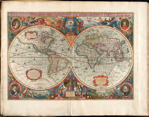
Hendrik Hondius, Nova totius terrarum orbis geographica ac hydrographica tabula, 1638. Boston Public Library, public domain.
Mercator developed the first atlases out of a colonial gaze, to codify all the earth's land masses and their people under what he called the "eye of history." Through their imagery, Mercator’s maps defined Europe as the sole source and determinant of meaning for the rest of the world.10 This 1638 Mercator-Hondius map flattens the globe into two sections, labeled the "New World" and the "Old World." These terms pitted Europe against the Americas in a binary opposition, portraying the latter as everything the former was not, positioning the cultures of the Americas as inferior to those of the colonizers. The bottom center depicts several women, who symbolize the major continents, offering up their resources to Europe, who sits on a throne. This atlas was not simply mapping natural space, but solidifying concepts of Eurocentrism that have lasted for centuries.11
How might we see reflections of this meaning formation in anatomical atlases? The atlas originated as a tool of classification that defines everything in the field it covers, as well as their relationships. While plotting out concepts of ideal body proportions, some Renaissance and Enlightenment anatomists used the “male sex” first, omitting equivalent female models from their treatise altogether. For Albinus and Wandelaar, “it went without saying that a perfect skeleton was perforce male.” They discarded any bones that made their illustration look round or slender, which they deemed too feminine.12 These anatomical atlases leaned on the Christian creation myth to reinforce the belief in an idealized universal body, created in the image of God, that is white, European, and male. We can see an example of a map-like diagram illustrating this in the Atlas Anatomico by Crisóstomo Alejandrino José Martínez y Sorli. All other forms of human anatomy become secondary or othered against the assumed default.
Echoes of Allegory
Each of these anatomists and illustrators had their own view of how to best portray scientific observations. We can see, however, how they built these viewpoints upon existing religious and philosophical worldviews, aesthetic conventions of their time, and in reaction to what came before. They mapped stories upon anatomy, in ways both stark and subtle, according to their understanding of hierarchies regarding religion, gender, race, and class. As with geographical atlases, there is an imperialist aspect to the anatomical gaze. It sets out to order, classify, and control. What echoes of Renaissance anatomical illustration can we still witness today? Those in power are increasingly attempting to regulate gender and reproduction along the lines of anatomy drawn by men who became cadavers themselves centuries ago.
Tracy Molis is an artist and adjunct professor in Visual Arts and Archaeology based in Brooklyn, NY. Her work, which includes paintings and archaeological illustrations, has been exhibited in galleries such as Nagel Draxler Kabinett, Berlin, Kai Matsumiya, New York, Clima, Milan, and Night Gallery, Los Angeles. She co-teaches the interdisciplinary course "Science and Art in Archaeological Illustration" with Archaeologist Zoë Crossland at Columbia University. Molis has worked with students at the New York City Archaeological Repository to create illustrations for artifacts in their collection and question the ways we represent the past.
Suggested Readings
Lorraine Daston and Peter Galison. “The Image of Objectivity.” Representations no. 40 (1992).
Martin Kemp. “Style and Non-Style in Anatomical Illustration: From Renaissance Humanism to Henry Gray.” Journal of Anatomy, vol. 216, no. 2 (2010).
Kemp, "M. Vesalius's Veracity." Nature 393 (1998).
José Rabasa. Inventing America: Spanish Historiography and the Formation of Eurocentrism (University of Oklahoma Press, 1993).
Citations
Kemp, Martin. “Style and Non-Style in Anatomical Illustration: From Renaissance Humanism to Henry Gray.” Journal of Anatomy, vol. 216, no. 2 (2010), 192-196.
Kemp, 199-200.
Taylor, Christina. “Spotlight 10: Muscles of the Back.” Dare to Know: Prints and Drawings in the Age of Enlightenment, Edouard Kopp, Elizabeth M. Rudy, and Kristel Smentek, eds. (Harvard: Harvard Art Museums, Yale University Press, 2022) 268-271.
Kemp, 198.
Daston, Lorraine and Peter Galison. “The Image of Objectivity.” Representations no. 40 (1992), 90-91.
Kemp, 200.
Kemp, 195.
Kemp, 200.
Kemp, 202
Rabasa, José. Inventing America: Spanish Historiography and the Formation of Eurocentrism (University of Oklahoma Press, 1993), 181.
Rabasa, 188-209.
Daston and Galison, 90.
Tracy Molis is an artist and adjunct professor in Visual Arts and Archaeology based in Brooklyn, NY. Her work, which includes paintings and archaeological illustrations, has been exhibited in galleries such as Nagel Draxler Kabinett, Berlin, Kai Matsumiya, New York, Clima, Milan, and Night Gallery, Los Angeles. She co-teaches the interdisciplinary course "Science and Art in Archaeological Illustration" with Archaeologist Zoë Crossland at Columbia University. Molis has worked with students at the New York City Archaeological Repository to create illustrations for artifacts in their collection and question the ways we represent the past.